FAQs

What is Cancer

The human body is made up of small building blocks called cells. All throughout life, these cells divide and re-divide to give rise to new cells. Normally, this process is tightly regulated in the body. Sometimes, due to certain factors, this process becomes unregulated. This unregulated cell division gives rise to cancers. There are many different types of cancers, and each one is treated differently.Diagnosis of cancer does not mean that you cannot be cured – many cancers if diagnosed early can be cured with proper treatment.

Breast Cancer

Breast tissue is made up of a bunch of glands called lobules, and ducts. Here, milk is produced, stored and carried through to the nipple during breastfeeding. Breast cancer starts in these ductal or lobular cells.
There are two main types of breast cancer:
  • Non-invasive breast cancer
  • Invasive breast cancer
Non- invasive breast cancer (Ductal Carcinoma in Situ – DCIS) This type of breast cancer means that the cancer cells are present in the milk ducts and have not spread into the breast tissue or other parts of the body. Invasive breast cancer In this type of cancer, the cancer cells are no longer limited to the ducts or lobules but have spread to the surrounding breast tissue and have potential to spread to other parts of the body.

Frequently Asked Questions

The best way to diagnose breast cancer is by a combination of clinical assessment, radiological tests, and pathological tests. This combination is known as “Triple Assessment” . If the doctor notices any suspicious findings during clinical examination, then the patient is advised to undergo tests such as mammography (for ladies above the age of 40 years), or ultrasound of the breasts (for ladies younger than 40 years), and pathological tests like fine needle aspiration cytology (FNAC), or core needle biopsy (CNB).

In a majority of the patients, these investigations are sufficient for the diagnosis of breast cancer. Rarely, additional tests (like MRI of breasts, PETCT scan, mammography guided/stereotactic/MRI guided biopsy, vacuum assisted breast biopsy (VABB), and occasionally open biopsy from the suspicious lump or nodule in the breast) may be required. In western countries, (occasionally in our country as well) due to regular screening, a large number of patients present with only mammography-detected abnormalities in the breast. These patients then need either mammography guided (or stereotactic) biopsy.

After the diagnosis of breast cancer, additional tests may be advised to assess the clinical stage of the disease and to plan the treatment.
Treatment of breast cancer is done by a team approach. The treatment plan includes surgery, chemotherapy, radiation therapy, hormonal therapy, and targeted therapy, individually or in a combination of two or more treatments. It means that all breast cancer patients do not need all parts of the treatment.

Breast cancer treatment plans are tailor made for individual patients. The treatment of an individual breast cancer patient is planned after considering many factors such as the patient’s age, activity level, nutritional status, stage of the disease, presence or absence of high risk factors in the pathological report, presence of any other medical illnesses, and the patient’s choice and compliance.

Unfortunately, in our country, the patient’s financial condition also plays an important role in the treatment plan, especially so if the treatment is carried out in private cancer centres.
Most women are able to return to a normal, productive lifestyle after the completion of breast cancer treatment. The recovery phase after surgery is usually not prolonged, and extensive periods of bed rest are not required. During and after chemotherapy and radiotherapy, except for a few days required to recover from the side effects of the treatment, the patient is usually able to continue to work as she had been before. Apart from this, she is also able to continue to play her various roles in the family and society. Women who continue to lead an active life with a positive frame of mind generally do not require a prolonged period of rehabilitation.
Breast cancer is the top cancer in women both in the developed and the developing world. The incidence of breast cancer is increasing in the developing world due to increased life expectancy, increased urbanization and adoption of western lifestyles. Although some risk reduction might be achieved with prevention, these strategies cannot eliminate the majority of breast cancers that develop in low- and middle-income countries, where breast cancer is diagnosed in very late stages. Therefore, early detection in order to improve breast cancer outcomes and survival remains the cornerstone of breast cancer control.

Limited resource settings with weak health delivery systems, where breast cancer incidence is relatively low and the majority of women are diagnosed in late stages, have the option to implement low-cost early diagnosis programmes based on awareness of early signs and symptoms, and prompt referral to diagnosis and treatment.

Population-based cancer screening is a much more complex public health undertaking than early diagnosis. It is usually cost-effective when done in the context of high-standard programmes that target all the population at risk in a given geographical area with a high specific cancer burden. Here, everyone who takes part is offered the same level of screening, diagnosis and treatment services.

So far the only breast cancer screening method that has proved to be effective is mammography screening. However, it is very costly and is cost-effective and feasible only in countries with good health infrastructure that can afford long-term, organized, population-based screening programmes. Low-cost screening approaches, such as clinical breast examination, could be implemented in limited resource settings when the necessary evidence from ongoing studies becomes available.

Many low- and middle-income countries that face the double burden of cervical and breast cancer need to implement combined cost-effective and affordable interventions to tackle these highly preventable diseases. The WHO promotes breast cancer control within the context of national cancer control programmes, and integrated non-communicable disease prevention and control.
Regular Breast Self examination once a month

Breast Cancer is curable if detected early.

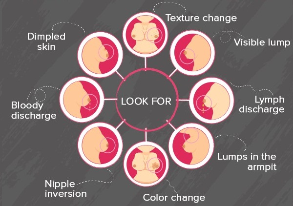
Consult Dr.Vaishali if you notice

  • Painless lump in the breast
  • Blood stained nipple discharge
  • Nipple retraction
  • Any other changes in the breast which persist for more than two weeks
Maintain a healthy weight by undertaking regular physical activity

Regular physical activity can help prevent breast cancer.


Balanced Diet

Eat a balanced diet rich in vegetables, fruits and proteins and low in fat


Talk

Talk to your breast surgeon about mammogram if you are above 40 years.

What to do if you find a lump in the breast

Not all lumps are cancerous. In younger age, just before and during monthly periods, one can feel lump(s) in the breast(s). A breast lump can be any one of the non- cancerous conditions like a breast cyst, a fibro-adenoma, an abscess, a sebaceous cyst under the breast skin, etc. A lump which is firm or hard to feel and usually does not go away after periods is suspicious of cancer and needs urgent attention. It is advisable to consult your breast surgeon to get assessed clinically. Some tests may be advised to understand the nature of the lump. Some of the tests which may be needed are Mammography, Ultrasound scan of the breasts, MRI of breasts, FNAC (Fine needle aspiration cytology), Core needle biopsy, etc.


What to do if you have family history of breast cancer?

There is a slightly high risk of developing breast cancer, if your first degree relative, i.e. mother, father, siblings etc. (male or female) has been diagnosed with breast or ovarian cancer. If more than one relative have been suffering from breast cancer or they have been diagnosed at a young age then the risk relatively increases. Nearly 5-10 % of breast cancers are due to inherited genetic abnormalities. A positive family history does not mean that one has to live in constant fear of developing the disease. Being aware of this fact is the most important thing. It is recommended that one should do regular self- breast examination once a month starting at the age of 18-20 years …..read more, get oneself examined by a breast surgeon if any change is noticed in the breast(s). The relatives (especially siblings and children) of patient who has been diagnosed with breast cancer at a young age (less than 40 years) should start screening 5 years before the age at which the patient was diagnosed. MRI of the breasts is usually recommended screening modality for those who are younger than 40 years. After 40 years of age annual mammography is recommended as a screening tool. This helps in diagnosis of breast cancer at an early stage.


What to do if you have nipple discharge?

Many women notice nipple discharge at various stages of life. Majority of the times the discharge is due to benign (non- cancerous) conditions of the breast. The discharge can vary in colour from watery, light yellow, milky, yellowish green or brown, or reddish, blood stained. It can be present in one or both sides at the same time. Nipple discharge can be seen coming out from a single duct opening or from multiple duct openings on the surface of the nipple. Many a times the nipple discharge is seen only after pressing below the nipple. Some women continue to have milky white discharge several months after discontinuing breast feeding. Common causes of nipple discharge are fibrocystic breast disease, duct ectasia, intra-ductal papilloma, etc. If the discharge is spontaneous (i.e. seen without pressing behind the nipple) or is seen to be coming from a single duct opening every time and is blood stained, then you are advised to consult your breast surgeon immediately. In addition to clinical assessment, you may be advised to undergo certain tests like ultrasound scan with high frequency probe, mammography, cytological examination of the discharge, or sometimes MRI of breasts, etc.